Many people choose LASIK to free themselves from glasses or contacts, expecting smooth recovery and lasting clarity. For most, that’s exactly what happens. But in a small number of cases, issues with the corneal flap can disrupt healing, blur vision, or cause discomfort.
The good news? LASIK flap complications can be corrected, and vision restored, but only with timely diagnosis and proper management.
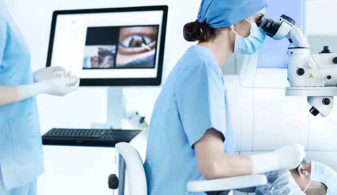
I’m Dr. Erica Darian-Smith, Ophthalmic Surgeon at Eagle Eye Surgeons in Sydney. I’ve managed many patients with flap-related concerns after LASIK, and I want to walk you through what they are, how we treat them, and what you can do to protect your vision.
Key Takeaways
- LASIK flap complications are uncommon, but early detection and treatment protect long-term vision.
- Sudden blur, pain, redness, or light sensitivity after LASIK can signal flap issues and need urgent care.
- Careful pre-operative screening, precise surgical technique, and strict post-LASIK care reduce the risk of flap complications.
- Most LASIK flap problems respond well to timely management, allowing patients to recover clear and stable vision.
- Alternatives like PRK or SMILE surgery avoid a corneal flap and may be safer options for higher-risk patients.
LASIK Flap Basics
What the flap is and why it matters
LASIK reshapes the cornea to correct vision. A thin corneal flap is created with a femtosecond laser. This flap is lifted so the underlying tissue can be shaped, then repositioned at the end of the procedure. A well-positioned flap supports smooth healing, stable vision, and comfort.
How flap complications are classified
- Intraoperative: happens during the flap creation or lifting process.
- Postoperative: During healing or years later.

Types of LASIK Flap Complications
Flap complications range from minor wrinkles to rare but vision-threatening events.
Intraoperative complications
Though uncommon, complications during flap creation can influence both the immediate surgery and long-term results.
- A buttonhole or free-cap flap occurs when the corneal flap is incomplete or completely detached, making it unsuitable for proper treatment.
- Irregular or decentered flaps can compromise the accuracy of laser reshaping, requiring careful adjustment or postponement.
- Femtosecond lasers can create an opaque bubble layer. It usually clears quickly but may reduce visibility during surgery.
Epithelial defects or interface debris can complicate early healing, highlighting the importance of careful surgical handling and intraoperative assessment to support a smooth recovery and optimal visual outcomes.
Postoperative complications
The period after surgery is when the flap is most vulnerable.
- Dislocation, partial or complete, may happen if the eye is rubbed or accidentally injured in the first days after LASIK.
- Flap striae, or wrinkles, can distort central vision, creating glare or ghosting.
- Epithelial ingrowth involves surface cells migrating under the flap, which can progress if untreated.
- Diffuse lamellar keratitis (DLK) develops under the flap and requires prompt steroid management.
- Central toxic keratitis (CTK) and infections are rare but serious, carrying the risk of permanent scarring if not identified and treated with urgency.
Other complications
Though very rare, some flap-related issues can have long-term effects on vision.
- Corneal ectasia occurs when the cornea progressively thins and bulges forward, reducing stability and clarity.
- Severe infectious keratitis can leave permanent scarring, leading to blurred or distorted vision.
- Irregular astigmatism, sometimes a by-product of flap issues or secondary scarring, reduces best corrected visual acuity and can limit the sharpness patients achieve even with glasses.
Signs and Symptoms Patients Should Recognise
Early symptoms can be harmless, but others mean trouble. The key is knowing which signals are normal and which need urgent review.
| Category | Typical Experiences | When It Signals a Problem |
| Normal Post-LASIK Symptoms | • Hazy vision in the first few days.
• Mild discomfort or scratchiness. • Dryness and light sensitivity. |
These are expected in early healing and usually improve with drops and rest. |
| Warning Signs of Flap Issues | • Sudden decline in clarity after initial improvement.
• Persistent redness or sharp pain. • Ghosting, double vision, or irregular surface with blinking. • Foreign-body sensation that doesn’t resolve. |
Suggests possible flap movement, inflammation, or early ingrowth. Requires urgent review. |
| Flap Striae Indicators | • Glare, halos, or starbursts around lights.
• Central blur or irregular astigmatism symptoms. • Reduced best-corrected vision if folds are central. |
Signs of wrinkling or folds in the flap, which can distort long-term clarity if untreated. |
Prevention Strategies Across the Patient Journey
Prevention starts before the laser is even switched on. Careful screening, precise technique, and disciplined aftercare reduce risks dramatically.
Preoperative screening
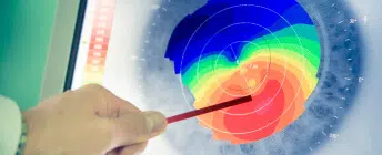
Thorough diagnostics protect against complications. This includes corneal tomography, epithelial mapping, dry-eye assessment, and ectasia risk scoring. Patients with higher flap risk may be advised to choose PRK, TransPRK, or SMILE.
Intraoperative best practices
Surgeons confirm suction, centration, and flap parameters before they work on the flap. Interface debris is avoided through controlled hydration and irrigation. Proper handling and precise repositioning reduce the chance of microfolds or instability.
Postoperative protection
Patients receive shields for sleep, instructions to avoid rubbing, and clear guidance on when to resume activities. Lubricating drops, antibiotics, and anti-inflammatory medications are prescribed. Follow-up visits confirm smooth healing and early detection of any issues.
How Specialists Diagnose and Grade Flap Issues
When problems arise, precise assessment is the first step. Modern imaging tools help identify flap position, folds, or early ingrowth.
In-clinic assessment
- Slit-lamp examination of the flap edge and interface.
- Fluorescein staining to highlight folds or irregularities.
- Anterior segment OCT or topography to map flap position, microstriae, or epithelial ingrowth.
Differentiating causes
Specialists distinguish between flap folds, epithelial basement changes, or tear-film instability. They must also separate inflammation (DLK) from infection, since management differs significantly.
Management Pathways for Specific Flap Issues
Treatment depends on the complication. Each pathway aims to restore clarity quickly and protect the long-term health of the cornea.
Flap dislocation
Prompt intervention is required. The flap is lifted, irrigated, smoothed, and precisely realigned. A bandage contact lens is placed, and topical antibiotics and steroids are prescribed. Sutures may be needed if instability persists.
Flap striae
Minor folds may be treated early with refloating and gentle stretching. More significant striae may require flap lifting, smoothing, and controlled drying. Persistent cases may need sutures to stabilise alignment.
Epithelial ingrowth
If peripheral and stable, observation may be safe. Central or progressive growth requires lifting the flap, scraping, and irrigating. Rare severe cases may require flap removal or conversion to PRK.
Inflammation and infection
- DLK is managed with staged corticosteroid therapy. This monitors intraocular pressure as needed.
- Infectious keratitis is treated urgently with culture-guided antibiotics to prevent permanent scarring.
Recovery, Prognosis, and Vision Outcomes
Most patients return to clear vision within days, but recovery is different for everyone.
Short-term recovery
Most LASIK patients notice clearer vision within days. Dryness can cause fluctuation, but this improves with consistent lubrication. Early management of flap complications supports rapid return to stable vision.
Long-term outlook
With timely treatment, outcomes are excellent. Patients who develop residual refractive error may be offered PRK, SMILE, or customised enhancement in the future.
Post-LASIK care timeline
| Stage | Care Instructions | Key Warnings |
| First Week | • Wear protective shields at night to avoid rubbing or pressure during sleep.
• Avoid rubbing, water exposure, heavy lifting, or straining. • Use prescribed drops exactly as directed to control inflammation, prevent infection, and support lubrication. |
• Sudden blur or vision loss.
• Sharp pain or strong foreign-body sensation. • Unusual light sensitivity or worsening redness. |
| Weeks to Months | • Taper anti-inflammatory medications under supervision.
• Continue lubrication to reduce dryness and improve comfort. • Resume activities gradually; delay high-impact sports until cleared. • Attend scheduled follow-ups to detect microstriae or epithelial ingrowth early. |
• Any new visual distortion or ghosting.
• Persistent dryness that does |
When Urgent Care Is Required
Recovery after LASIK is usually smooth, but certain signs mean it’s time to call your ophthalmologist right away.
Sudden blur, sharp pain, or redness that spreads should never be ignored, as they can point to flap displacement, infection, or inflammation. Light sensitivity that worsens rather than improves may also signal trouble, as can any unusual discharge.
Patients sometimes describe a sensation that the surface is uneven or that the flap has shifted. This is another red flag. Prompt examination allows specialists to reposition, treat, or medicate as needed, protecting long-term vision and, which may prevent permanent complications.
LASIK Versus Alternatives When Flap Risk Is High
Surface procedures such as PRK or TransPRK avoid a flap entirely, while SMILE uses a small incision. These methods lower flap-related risk but come with trade-offs in recovery and comfort. PRK takes longer to heal, while SMILE and LASIK both offer faster visual recovery. Careful screening is important to find the best method for the patient.
Frequently Asked Questions
Patients often have the same concerns: how stable is the flap, how common are complications, and can problems be fixed? The FAQ section addresses these directly.
How do I know if I messed up my flap after LASIK?
You might notice sudden blurred or distorted vision, discomfort, redness, or a gritty sensation. If your vision clarity drops after initially improving, or if pain increases, it’s important to see your eye surgeon promptly for a proper examination.
How common is flap dislocation after LASIK?
Flap dislocation is rare, occurring in less than 1% of patients. The risk is greatest in the first week after surgery, when the flap is most fragile, but significant trauma can sometimes dislodge it even years later.
Does the flap ever heal after LASIK?
The flap bonds securely to the cornea over time but never regains the full natural strength of unoperated tissue. For everyday activities it is stable, yet very forceful rubbing or direct trauma can still disturb it.
How to know if a flap is dislodged?
Signs include sudden vision changes, ghosting, halos, discomfort, or feeling that the eye surface is uneven. Only an eye doctor can confirm dislocation using a slit-lamp exam, so any suspicion should be checked urgently.
What are the complications of LASIK flaps?
Possible complications include flap dislocation, folds or striae, epithelial ingrowth, inflammation, or infection. Most are uncommon, and with early detection and treatment, long-term visual outcomes remain excellent.
Can flap dislocation be fixed?
Yes. Surgeons can lift, clean, and reposition the flap, often smoothing out folds and placing a bandage lens. When treated quickly, vision usually returns to normal with minimal long-term impact.
What are the symptoms of flap striae?
Flap striae can cause blurred vision, glare, halos, or starbursts, especially noticeable at night. Patients may also feel their vision is distorted or slightly “wrinkled,” reducing clarity if folds affect the central cornea.
What happens if a LASIK flap is lost?
Flap loss is extremely rare. If it occurs, doctors may use surface laser techniques such as PRK to restore vision. Outcomes vary depending on how quickly treatment is received, but useful vision is often preserved.
How hard is it to dislodge a LASIK flap?
In the first few days, even mild rubbing or bumping can move the flap. After healing, everyday life is safe, though significant trauma—such as an eye injury during sport—could still dislodge it years later.
Final Thoughts
LASIK remains one of the most effective ways to reduce reliance on glasses or contact lenses. Flap complications are uncommon, and with modern diagnostics and prompt care, most patients recover strong, stable vision.
If you’re considering LASIK or have concerns after surgery, an ophthalmology consultation offers clarity and reassurance. At Eagle Eye Surgeons, we provide the expertise and support needed to protect your sight for the long term.

– Fellow of the Royal Australian and New Zealand College of Ophthalmologists (FRANZCO)
– Fellow of World College of Refractive Surgery and Visual Sciences (FWCRS)
– GradDipGraduate Diploma in Cataract and Refractive Surgery (University of Sydney)
– Master of Medicine (MMed, Ophthalmic Sciences, University of Sydney)
– Bachelor of Medicine and Surgery (MBBS, University of Tasmania)
Dr. Erica was a recipient of the 2022 ASCRS Foundation Resident Excellence Award. In 2019, she was awarded the RANZCO Filipic Greer Medal for overall excellence in performance at the RANZCO Ophthalmic Pathology examination. Most recently, she was awarded the Royal Australian and New Zealand College of Ophthalmologists (RANZCO) Trevalyn-Smith Travelling Scholarship to subsidize overseas study for Fellows.
As an accomplished researcher Dr. Erica’s work has been published widely in high quality medical journals, including the American Journal of Ophthalmology, the Journal of Cataract and Refractive Surgery, the European Journal of Ophthalmology and Clinical and Experimental Ophthalmology. Erica has also written a book chapter and has had the opportunity to present her research at various international and national conferences. Dr. Erica is appointed as a Clinical Lecturer in the Discipline of Ophthalmology at the University of Sydney, Save Sight Institute and regularly contributes to ongoing teaching in her area of subspeciality.