Vitreoretinal Surgery: Retinal Detachment, Macular Hole, and Epiretinal Membrane
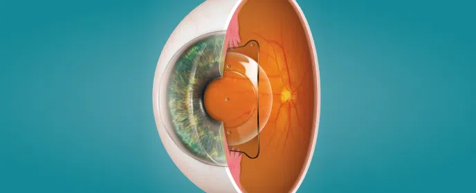
The retina is a vital part of our eyes that plays a significant role in our vision. It is a thin layer of tissue located at the back of the eye that captures light and sends signals to the brain, allowing us to perceive the world around us. It can be thought of as similar to the film in an analogue camera, or the sensor in a digital camera. There are many conditions that can affect the health and functionality of the retina, leading to vision problems. Three common causes are: retinal detachment, macular hole, and epiretinal membrane.
What do you need help with?
Vitreoretinal surgery
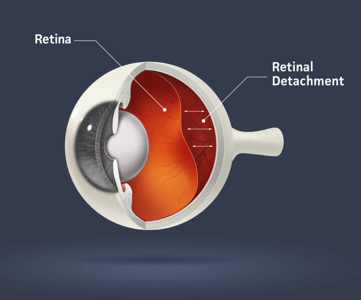
Retinal Detachment
Retinal detachment occurs when the retina becomes separated from the underlying tissue that supports and nourishes it. This is often considered a medical emergency as it can lead to permanent vision loss if left untreated. There are three types of retinal detachment: rhegmatogenous, tractional, and exudative.
- Rhegmatogenous retinal detachment is the most common type and is caused by a hole or tear in the retina, allowing fluid to seep underneath and separate it from the underlying tissue.
- Tractional retinal detachment occurs when scar tissue on the retina’s surface pulls it away from the supporting tissue.
- Exudative retinal detachment is caused by fluid accumulation beneath the retina due to inflammation or injury.
Understanding Retinal Detachment: Causes, Symptoms, and Treatment Options

Causes and risk factors of retinal detachment
Retinal detachments can be caused by various factors. These include trauma or injury to the eye, severe nearsightedness, previous eye surgery, family history of retinal detachment, and certain predisposing eye conditions. Age and gender can also play a role. Retinal detachment is more common in individuals over the age of 40 and in men.

Symptoms and diagnosis of retinal detachment
The symptoms of retinal detachment can vary depending on the type and severity of the detachment. Common symptoms include sudden onset of floaters, flashes of light, a shadow or curtain-like effect in the peripheral vision, and a decrease in vision. If you experience any of these symptoms, it is essential to seek immediate medical attention from an eye care professional. They will perform a comprehensive eye examination, which may include a dilated eye exam, ultrasound imaging, or optical coherence tomography (OCT), to diagnose retinal detachment.

Treatment options for retinal detachment
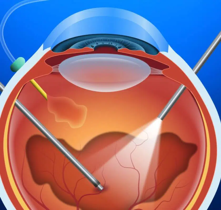
The treatment for retinal detachment depends on the type and severity of the detachment. In most cases, surgery is required to reattach the retina to the underlying tissue. The two main surgical procedures used to treat retinal detachment are scleral buckle and vitrectomy. Scleral buckle involves placing a silicone band around the eye to provide support and bring the detached retina back into place. Vitrectomy, on the other hand, involves removing the vitreous gel from the eye and replacing it with a gas bubble or silicone oil to push the retina against the supporting tissue. Retinal detachment is a complex condition, and a small number of cases (10-15%) require multiple surgical procedures to successfully reattach the retina. Dr Lee is a fellowship trained vitreoretinal surgeon, and he will determine the most suitable treatment option for you.
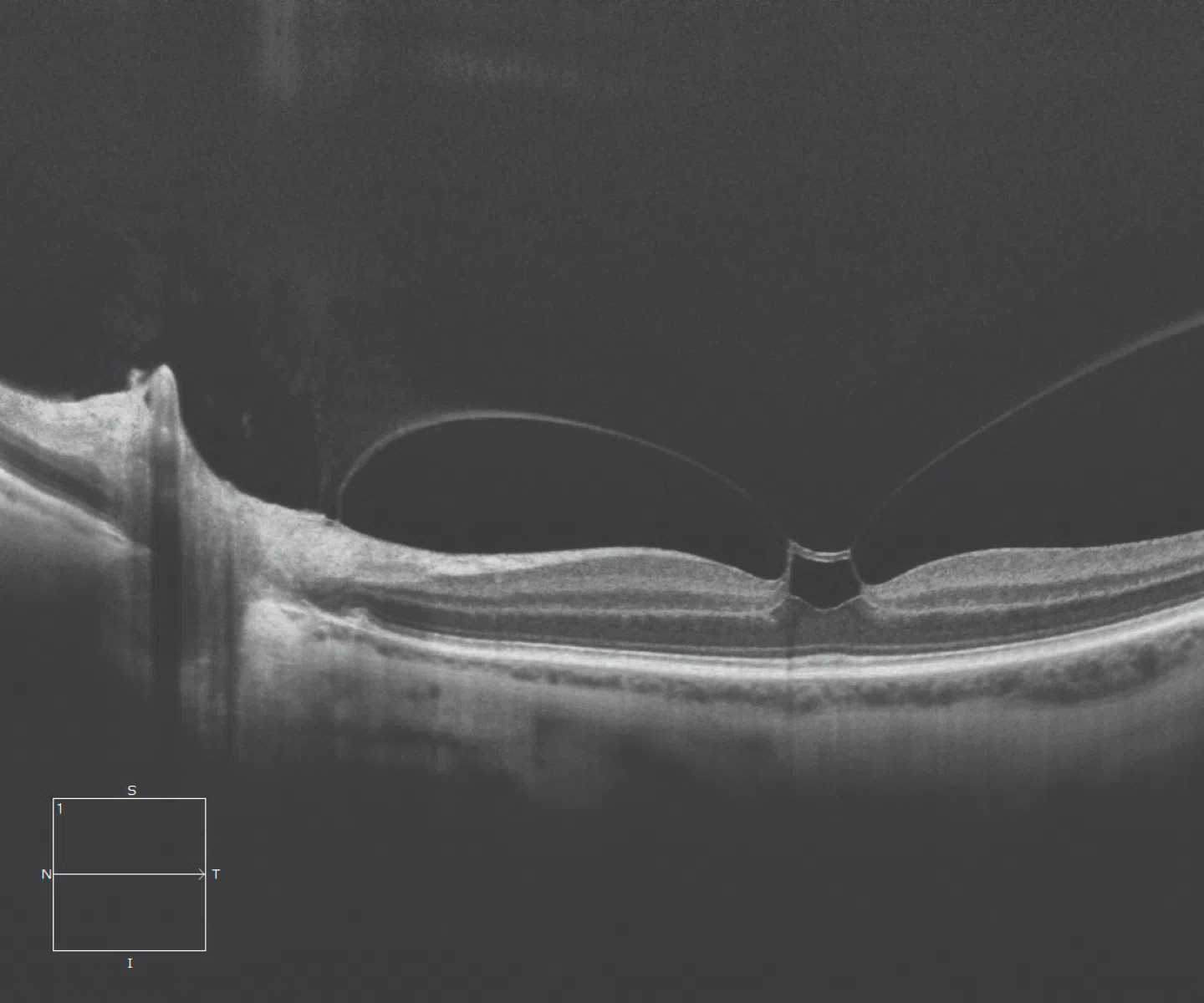
Macular Hole
A macular hole is a condition that affects the center of the retina, known as the macula. The macula is responsible for sharp, central vision, which allows us to read, drive, and recognize faces. When a hole forms in the macula, it can significantly impact our ability to perform these tasks. Macular holes are often associated with age-related changes in the eye. They primarily affect individuals over the age of 60.
Understanding Macular Hole: Causes, Symptoms and Treatment Options
Causes and risk factors of macular holes
The primary cause of macular holes is the natural aging process, which leads to the shrinkage of the vitreous gel within the eye. As the vitreous gel shrinks, it can pull on the macula, creating a hole. Other risk factors for macular holes include trauma to the eye, certain eye conditions such as diabetic retinopathy or high myopia, and previous eye surgery. Women are more likely to developing macular holes than men.
Symptoms and diagnosis of macular holes
The symptoms of macular holes can vary depending on the stage and size of the hole. In the early stages, you may experience blurry or distorted central vision, difficulty reading or performing close-up tasks, and a dark spot in the center of your vision. As the hole progresses, these symptoms may worsen. If you notice any changes in your central vision, it is important to consult with an eye care professional. They will perform a comprehensive eye examination, including a dilated eye exam and optical coherence tomography (OCT), to diagnose a macular hole.
Treatment options for macular holes
The treatment for macular holes typically involves surgery to close the hole and restore vision. The most common surgical procedure used to treat macular holes is called a vitrectomy. During this procedure, the vitreous gel is removed from the eye, traction on the edges of the hole is relieved, and the hole is closed with a gas bubble. Over time, the gas bubble is absorbed, and the eye fills with natural fluid. After surgery, you will be required to maintain a face-down position for a certain period to optimize the healing process. Surgery for small macular holes has a first operation success rate of 90-95%. Large macular holes and those that have been present for a long time have a lower first operation success rate (80% or less). Cases of recurrent or persistent macular holes require treatment with more advanced surgical procedures including flap-based repairs, amniotic membrane grafting, and posterior pole detachment techniques. Dr Mitch is a fellowship trained vitreoretinal surgeon who has expertise in all forms of macular hole surgery, and he will determine the most suitable treatment option for you.

Epiretinal Membrane
An epiretinal membrane, also known as a macular pucker, is a thin layer of scar tissue that forms on the surface of the macula. This scar tissue can cause the macula to wrinkle or distort, leading to blurred or distorted vision. Epiretinal membranes are often associated with age-related changes in the eye and are more common in individuals over the age of 50.
Understanding Epiretinal Membrane: Causes, Symptoms, and Treatment Options
Causes and risk factors of epiretinal membrane
The exact cause of epiretinal membranes is unknown. They are thought to develop as a result of changes in the vitreous gel and the subsequent formation of scar tissue at the back of the eye. Risk factors for epiretinal membranes include age, previous eye surgery or trauma, certain eye conditions such as retinal tears or detachments, and certain medical conditions such as diabetes.
Symptoms and diagnosis of epiretinal membrane
The symptoms of epiretinal membranes can vary depending on the severity of the condition. Common symptoms include blurry or distorted central vision, difficulty reading or recognizing faces, and the perception of straight lines as wavy or bent. If you experience any of these symptoms, it is important to schedule an appointment with an eye care professional. They will perform a comprehensive eye examination, including a dilated eye exam and optical coherence tomography (OCT), to diagnose an epiretinal membrane.
Treatment options for epiretinal membrane
In mild cases of epiretinal membranes, no treatment may be necessary. Regular monitoring of the condition may be recommended. If the symptoms significantly affect your daily life, surgical intervention may be needed. The most common surgical procedure used to treat epiretinal membranes is a vitrectomy, similar to the procedure used for macular holes. During the surgery, the scar tissue is carefully removed, allowing the macula to flatten and restore normal vision. Dr Mitch is a fellowship trained vitreoretinal surgeon, and he will determine the most suitable treatment option for you.

Prevention and management of retinal conditions
While some retinal conditions cannot be completely prevented, there are certain steps you can take to reduce your risk and manage these conditions effectively. Regular eye examinations are crucial for detecting any early signs of retinal conditions and allowing for prompt treatment. Protecting your eyes from trauma and injury by wearing appropriate protective eyewear is also important. Maintaining a healthy lifestyle, including a balanced diet rich in antioxidants, not smoking, and managing systemic conditions such as diabetes or hypertension, can help support the overall health of your eyes.
Retinal conditions, such as retinal detachment, macular hole, and epiretinal membrane, can have a significant impact on your vision and overall quality of life. At Eagle Eye Surgeons’ our subspecialty vitreoretinal surgeon, Dr Mitchell Lee, is a specialist at treating these conditions.

Meet Dr. Mitch Lee
Ophthalmic Surgeon, Vitreoretinal, Medical Retina, Cataract, Complex Anterior Segment, Refractive and General Ophthalmology
Dr Mitch is Eagle Eye Surgeons’ vitreoretinal fellowship trained surgeon. He trained at Prince of Wales Hospital in Sydney, before undertaking subspecialty fellowship training in vitreoretinal surgery at Westmead hospital. Dr Mitch is highly trained in medical and surgical treatments for patients with vitreoretinal conditions.

Book an appointment now
Macular Degeneration
Understanding Macular Degeneration: Causes, Symptoms, and Treatment Options
Macular degeneration is a common eye condition that affects millions of people worldwide. It is a leading cause of vision loss, particularly in individuals over the age of 50. By understanding this condition, individuals can take proactive steps towards managing their eye health and preserving their vision.
What is Macular Degeneration?
Macular degeneration, also known as age-related macular degeneration (AMD), is a progressive eye disease that affects the macula, the central portion of the retina responsible for sharp, central vision. The macula allows us to see fine details and perform tasks such as reading, driving, and recognizing faces. When the macula deteriorates, it can lead to significant vision loss and impairment.

Understanding Macular Degeneration: Causes, Symptoms, and Treatment Options
Causes of Macular Degeneration
The exact causes of macular degeneration are not fully understood. Several factors are known to contribute to its development. Age is the most significant risk factor, with macular degeneration being more common in individuals over the age of 50. Genetics also play a role, as certain gene variations have been associated with an increased risk of developing the condition. Smoking, obesity, high blood pressure, and a poor diet can increase the likelihood of developing macular degeneration.
Understanding the Different Types of Macular Degeneration – Wet and Dry
Macular degeneration can be categorized into two main types: wet and dry. Dry macular degeneration is the most common form. It occurs when the cells in the macula gradually break down, ultimately leading to a gradual loss of central vision. Wet macular degeneration occurs when abnormal blood vessels grow underneath the retina and leak fluid or blood, causing rapid and significant vision loss. It always occurs in conjunction with dry AMD. Not all patients with dry AMD will develop the wet form.
Symptoms of Macular Degeneration
The symptoms of macular degeneration may vary depending on the type and stage of the condition. In the early stages, individuals affected may not experience any noticeable symptoms. However, as the disease progresses, common symptoms may include blurred or distorted vision, a dark or empty area in the center of vision, difficulty recognizing faces, and a decrease in color perception. It is essential to be aware of these symptoms and seek prompt medical attention if any changes in vision occur.

Diagnosing Macular Degeneration
Diagnosing macular degeneration typically involves a comprehensive eye examination conducted by an ophthalmologist or optometrist. The eye care professional will evaluate the patient’s medical history, perform a visual acuity test, and conduct a dilated eye examination to examine the retina and macula. Additional tests, such as optical coherence tomography (OCT), Ultra-widefield fundus imaging and/or fluorescein angiography, may be performed to assess the extent of the damage and determine the type of macular degeneration.
Treatment Options for Macular Degeneration
While there is currently no cure for macular degeneration, several treatment options are available to manage the condition and slow its progression. The most common treatment approaches include intravitreal injections, medications, surgical procedures, and lifestyle changes.

Intravitreal Injections
Intravitreal injections involve the administration of medication directly into the eye. This approach is commonly used for treating wet macular degeneration. Anti-vascular endothelial growth factor (anti-VEGF) drugs are injected into the vitreous, the gel-like substance in the center of the eye. These drugs help reduce abnormal blood vessel growth and leakage, thereby preserving vision and preventing further damage. Treatment with anti-VEGF medications is highly effective, but it does not cure the condition. Injections need to be continued long term to maintain control over the disease and slow or halt its progression. Our Eagle Eye Surgeons will use their expertise to determine the best treatment schedule for your individual case.

Medications for Macular Degeneration
Supplements such as Macuvision Plus, containing antioxidant nutrients and minerals including Vitamin E, Vitamin C, Zinc, Lutein and Zeaxanthin may be recommended for certain types of macular degeneration. It is important to note that medications will not reverse the damage caused by macular degeneration, but may slow down its progression and help to preserve existing vision.

Surgical Procedures for Macular Degeneration
In some cases, surgical interventions may be necessary to manage macular degeneration. One such procedure is a vitrectomy, which involves the removal of the vitreous gel and treatment of bleeding beneath the retina. This procedure can help improve vision and reduce the risk of further vision loss. Dr Lee is a fellowship trained vitreoretinal surgeon who specializes in managing all medical and surgical aspects of macular degeneration.
Lifestyle Changes and Self-Care for Managing Macular Degeneration
In addition to medical interventions, certain lifestyle changes and self-care practices can play a significant role in managing macular degeneration. These include:
Eating
Eating a nutritious diet rich in antioxidants, vitamins, and minerals.
Quitting
Quitting smoking, as smoking has been strongly linked to an increased risk of macular degeneration.
Wearing
Wearing sunglasses that provide protection against harmful ultraviolet (UV) rays.
Regular exercise
Regular exercise to improve overall health and blood circulation.
Using assistive devices
Using assistive devices and technologies, such as magnifiers and voice-controlled devices, to enhance independence and daily functioning.
Diabetic Eye Disease

What is Diabetic Eye Disease?
Diabetic eye disease, also known as diabetic retinopathy, is a serious condition that affects the eyes of individuals with diabetes. It is caused by damage to the blood vessels in the retina, the light-sensitive tissue at the back of the eye.
Understanding Diabetic Eye Disease: Causes, Symptoms, and Treatment Options
Causes of diabetic eye disease
The primary cause of diabetic eye disease is uncontrolled diabetes. When blood sugar levels are consistently high, it can cause damage to the blood vessels throughout the body, including those in the eyes. Over time, high blood sugar levels continue to weaken and damage these retinal blood vessels, leading to vision problems and, in severe cases, blindness.

Understanding the symptoms of diabetic eye disease
Diabetic eye disease often develops gradually and may not cause noticeable symptoms in the early stages. However, as the condition progresses, individuals may experience symptoms such as blurred vision, floaters (tiny specks or spots that appear to float across the field of vision), difficulty seeing at night, and changes in color perception. It is important to note that these symptoms can also be indicative of other eye conditions, so it is crucial to consult an eye care professional for a proper diagnosis.

The importance of early detection and treatment
Early detection and treatment of diabetic eye disease are crucial for preserving vision. During an eye exam, your eye care professional will dilate the pupils with eye drops to get a clear view of the retina. They may also perform additional tests, such as Ocular Coherence Tomography (OCT) or Ultra-widefield fundus imaging to evaluate the retina. Early detection allows for timely intervention and treatment to prevent further damage to the eyes.
Regular eye examinations are essential for early detection of diabetic eye disease. If you have diabetes, it is recommended to have a comprehensive eye exam at least once a year. This will enable your eye doctor to identify any signs of retinopathy and initiate timely treatment to prevent vision loss.
Treatment options for diabetic eye disease
The treatment options for diabetic eye disease vary depending on the severity of the condition. In the early stages, when there is minimal or no vision loss, lifestyle modifications and close monitoring of blood sugar levels may be sufficient to manage the disease. As the disease progresses, more advanced treatments may be necessary. These can include laser treatments and injections of specialised anti-VEGF medications into the eye to reduce inflammation and swelling. In severe cases surgery can be necessary to remove scar tissue or repair diabetes-associated retinal detachments.
Preventing diabetic eye disease
Prevention plays a crucial role in managing diabetic eye disease. Maintaining good control of blood sugar levels, blood pressure, and cholesterol can significantly reduce the risk of developing the condition. Regular exercise, a healthy diet, and regular eye examinations are also essential in preventing the onset and progression of diabetic eye disease. It is important for people with diabetes to work closely with their healthcare team to develop a comprehensive plan for managing their overall health, including regular eye care.

Lifestyle changes to support eye health
In addition to medical interventions, certain lifestyle changes can help support eye health for individuals with diabetic eye disease. Quitting smoking is vital, as smoking can further damage blood vessels and exacerbate eye-related complications. Protecting the eyes from harmful UV rays by wearing sunglasses and using protective eyewear when necessary is also important. Maintaining a healthy weight, engaging in regular physical activity, and managing stress levels can all contribute to overall eye health and well-being.
Diabetic eye disease is a serious condition that can have a significant impact on vision and overall quality of life. If you have diabetes it is important to have regular eye examinations to detect the condition early. Our vitreoretinal surgeon, Dr Mitchell Lee, is experienced at treating all aspects of diabetic eye disease.